A Thoroughbred racehorse in full stride during a race at Churchill Downs, the historic racetrack in Louisville, Kentucky. The horse is mid-gallop, with all four hooves off the ground, demonstrating peak speed and muscular exertion. The ability of a racehorse to maintain a steady state internal environment while racing is key to a winning performance.
Learning Objectives
Define homeostasis and explain its importance in maintaining a stable internal environment.
List and explain the three major components of a biological control system: receptor, control center, and effector.
Differentiate between negative and positive feedback mechanisms with examples.
Discuss how exercise challenges homeostatic control systems and the body’s response to these challenges.
Describe how exercise training improves homeostatic control through physiological adaptations.
Explain the process of protein synthesis in response to exercise.
Identify the different cell-signaling mechanisms involved in cellular adaptations.
Define stress proteins and their function in protecting cells from damage.
Analyze how different types of exercise (resistance vs. endurance) lead to specific adaptations in muscle cells.
Evaluate the effects of environmental stressors on homeostatic control and the concept of acclimation.
Control of the Internal Environment
For over a century, physiologists have recognized that the “milieu intérieur” (internal environment) of the human body remains constant despite changing external conditions. Claude Bernard (1813-1878), a French physiologist and one of the founding fathers of physiology, emphasized the importance of a stable internal environment. Bernard discovered that the liver could synthesize glucose from blood-derived products like lactate and that the nervous system controls vasomotor responses, which can dilate or constrict blood vessels. Maintaining a stable internal environment is crucial for health. This chapter introduces exercise as a challenge to homeostatic control, reviews homeostatic control systems, and explains how adaptations affect these systems. Understanding the challenges exercise poses to homeostasis can help appreciate how exercise-induced adaptations protect the body from future stressors.
Exercise: A Challenge of Homeostatic Control
Exercise significantly challenges homeostatic control. Variables such as pH, core temperature, heart rate, blood pressure, ventilation, and hormone concentrations can deviate greatly from resting values during exercise. Homeostasis, a term coined by Walter Cannon in 1932, refers to the maintenance of a relatively constant internal environment, describing a dynamic balance that keeps the body within livable limits during rest. Steady state, while also describing a stable internal environment, refers to conditions where physiological variables are elevated or decreased from resting values. Steady state exercise involves maintaining constant but elevated levels of variables like heart rate, ventilation rate, body temperature, oxygen consumption, blood pressure, hormones, and blood glucose concentration are constant but elevated from rest.
Although homeostasis and steady state imply stability, the internal environment is not constant. These states are maintained through dynamic balance by control systems that make small adjustments to keep physiological variables around a “set” value. For example, blood pressure is a critical variable to control, as it ensures oxygen delivery to tissues. Resting blood pressure can oscillate between 92 and 94 mmHg, with an average arterial pressure around 93 mmHg[1]. During steady state exercise, blood pressure increases from rest and varies depending on individual training and exercise intensity (e.g. speed, resistance, load) of the mode of exercise. If exercise intensity remains constant, small oscillations in blood pressure occur but stay close to the mean steady state value (Figure 2.1). These oscillations result from highly regulated biological control systems that provide feedback to adjust blood pressure when it deviates from the set value[2].
Figure 2.1 Resting arterial blood pressures across time. Notice that the small oscillations revolve around the central mean pressure.
Homeostatic Regulation
Biological Control Systems
The human body contains thousands of control systems that maintain homeostasis. These systems range from intricate mechanisms within single cells to those regulating entire organs. They also manage interactions between organ systems. For example, the respiratory system, in conjunction with the nervous system, regulates carbon dioxide levels produced during metabolism, especially during exercise. A typical biological control system comprises three main components: the receptor, control center, and effector. The receptor, a specialized sensor, measures the current state of the system. The control center compares this value to the desired “set” value. If a discrepancy is detected, the effector modifies the parameter to correct the disturbance. This process often involves negative feedback, where the response reduces the original stimulus. The three components of a biological control system are:
1. Receptor: obtains current information
2. Control center: compares current value and compares it to a desired value
3. Effector: modifies some parameter
Negative Feedback
Most biological control systems operate via negative feedback, where the system’s response opposes the original stimulus. For instance, consider the regulation of blood glucose after eating shown in Figure 2.2. Following a high-carbohydrate meal, glucose enters the bloodstream, causing hyperglycemia (blood glucose levels above 100 mg/100 ml)[3]. Normal fasting blood glucose levels range from 80-90 mg/100 ml. In response, the pancreas releases insulin, which binds to cell receptors and facilitates glucose uptake into cells, thereby lowering blood glucose levels. This mechanism restores homeostasis by reducing blood glucose to normal levels. Negative feedback is so named because the control system’s response is in the opposite direction of the initial stimulus.
Figure 2.2 Illustration of the negative feedback that is used to regulate blood glucose levels after a high-carbohydrate meal. See text for details of how this system operates.
During exercise, several physiological control systems operate via negative feedback to maintain homeostasis. These include the regulation of carbon dioxide and oxygen concentrations, arterial blood pressure, body temperature, heart rate, and electrolyte balance. A common household thermostat, as illustrated in Figure 2.3, also employs a negative feedback system. The thermostat can be set to a desired temperature, such as 72°F, and is connected to the heating unit of a dwelling. If the indoor temperature drops below this setting, the thermostat activates the furnace to produce heat until the desired temperature is reached. Once the thermostat measures the set temperature (72°F), the furnace turns off. If the temperature drops again, the thermostat reactivates the furnace. In this example, the furnace’s response (producing heat) is opposite to the initial stimulus (a decrease in temperature).
In general, when a factor becomes excessive or deficient, a control system initiates negative feedback to make adjustments that return the factor toward a certain mean value, thereby maintaining homeostasis.
Figure 2.3 A common household thermostat set to 72°F. A thermostat also works by way of negative feedback.
Positive Feedback
Positive feedbackmechanisms amplify the original stimulus, meaning the response moves in the same direction as the stimulus. This can sometimes lead to vicious cycles and instability, potentially causing harm or even death. For example, consider body temperature regulation during endurance exercise in hot and humid conditions. Normally controlled by negative feedback, body temperature can rise from its normal level of 98.6°F to 102°F or 103°F (37°C to 40°C) due to heat produced by muscle contractions. In hot and humid environments, the body struggles to dissipate this heat, causing temperatures to potentially rise to 106°F to 108°F (41°C to 42°C). Such high temperatures can damage cells and lead to heatstroke, a dangerous positive feedback loop where the body’s temperature-regulating mechanisms fail, and the elevated temperature accelerates chemical reactions, producing even more heat[4]. Without intervention, this cycle can be fatal.
However, positive feedback can be beneficial in certain situations, such as childbirth, blood clotting, and nerve signal generation. For instance, to generate a nerve signal during exercise, a motor neuron must be stimulated, causing sodium ions to leak into the cell. This changes the membrane potential, opening more sodium channels and allowing more sodium to enter, eventually triggering an action potential and nerve transmission to stimulate muscle contraction. This positive feedback process enables neurons to function within numerous negative feedback systems, maintaining overall homeostasis through interconnected control systems.
Gain of the control system
The effectiveness of a control system in maintaining homeostasis through negative feedback varies, described by the term “gain.” Gain is a term to describe the precision with which a control system maintains homeostasis. Systems with high gain maintain homeostasis more precisely than those with low gain. Gain is determined by the system’s ability to correct disturbances and the error involved in preventing changes. The gain of a system can be calculated using the following formula:
This formula quantifies the system’s precision in maintaining stable conditions.
Exercise Improves Homeostatic Control
Exercise challenges the body’s homeostatic control systems by potentially disrupting variables such as core temperature, acid-base balance, and oxygen and carbon dioxide levels. During sub-maximal exercise in a cool environment, control systems can maintain a steady state. However, prolonged or intense exercise in hot or humid conditions can overwhelm these systems, leading to premature fatigue or cessation of exercise. Heavy exercise can cause disturbances too great for even the most effective control systems to manage, preventing a steady state. Exercise training can improve performance under these conditions by enhancing homeostatic control.
Exercise training stimulates physiological adaptations in affected organ systems, improving homeostatic control. Adaptations involve changes in the structure and function of cells, tissues, or organ systems, enhancing the ability to maintain a steady state during stress, such as exercise, and allowing the body to return to homeostasis quickly afterward. The principle of specificity states that exercise adaptations are specific to the muscles involved, the muscle fiber types recruited, and the energy systems used. For example, aerobic exercise leads to adaptations in oxygen transport (increased synthesis of myoglobin) and utilization (more mitochondria) mechanisms, while anaerobic resistance training increases proteins associated with force production (actin and myosin) and anaerobic energy creation (creatine kinase). These adaptations are specific to the type of training and develop over weeks. Exercise improves homeostatic control through adaptation.
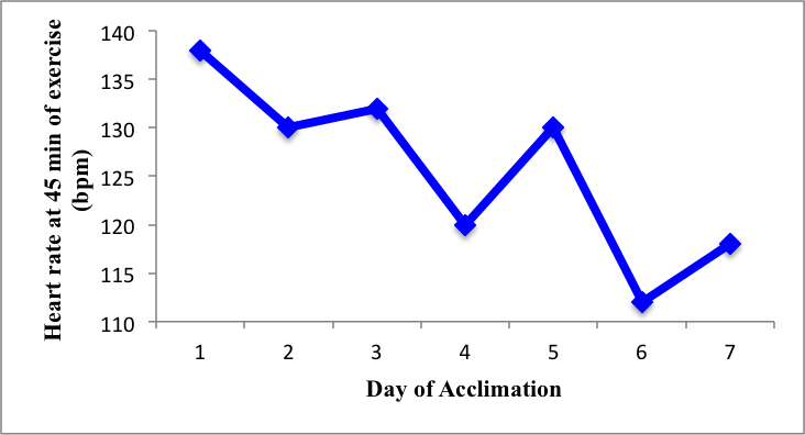
Exposure to environmental stressors also causes adaptations, known as acclimation, which occur after repeated or chronic exposure to conditions like heat, altitude, cold, deep-sea diving, or space flight. These adaptations are studied in Environmental Exercise Physiology and are of interest to organizations like the National Aeronautics and Space Administration (NASA). Figure 2.4 shows the relationship of heart rate (bpm) at 45 minutes of cycling exercise (50 W) throughout a 7-day acclimation protocol in 40°C. Note that the heart rate is lower during day 7 of the protocol (~118 bpm) when compared to the same time during day 1 (~138). This decrease in heart rate following several days of exercise in the heat demonstrates acclimation of the cardiovascular and associated systems[5]. Acclimation results in the improved function of an existing homeostatic system. When environmental conditions are artificially created, such as in altitude chambers, the resulting adaptations are called acclimatization. Training-induced adaptations enhance performance by improving the body’s ability to maintain homeostasis.
Figure 2.4 The relationship between heart rate at the end of 45 minutes of cycling (50 W) in 40°C and days of acclimation.
Cellular Adaptations
Exercise training induces cellular adaptations that enhance the function of existing homeostatic systems. Cell signaling refers to the communication processes that occur within or between cells, allowing them to coordinate activities. There are five major cell-signaling mechanisms involved in adaptation and homeostasis:
Intracrine signaling. Chemical messengers within a cell trigger a response in the same cell.
Juxtacrine signaling. Adjacent cells communicate through transmembrane protein junctions that allow a chemical messenger to travel from one cell to the neighboring cell.
Autocrine signaling. A cell secretes a chemical messenger into the extracellular fluid, but the receptor for this messenger is on the membrane of the same cell that produced it.
Paracrine signaling. A cell communicates with nearby cells by secreting a chemical messenger into the extracellular fluid.
Endocrine signaling. Cells secrete hormones into the bloodstream, affecting downstream cells that have specific receptors for these hormones.
Exercise Stimulates Protein Synthesis
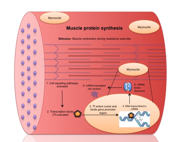
Regular exercise induces specific cellular adaptations depending on the type of training. Resistance training and endurance training lead to different adaptations, coordinated by cell signaling mechanisms. The mechanical and metabolic stimuli of exercise activate signaling pathways, leading to protein synthesis and subsequent adaptations within muscle cells. Understanding the process of protein synthesis is crucial, as exercise stimulates structural and metabolic changes in this manner. The process of protein synthesis induced by exercise involves the following steps and is shown in Figure 2.5:
1. The mechanical mechanisms and metabolic stresses of exercise activate a cell-signaling pathway.
2. The pathway activates a transcription factor, which enters the cell nucleus.
3. In the nucleus the transcription factor binds to a gene promoter region, initiating DNA transcription.
4. DNA is transcribed to messenger RNA (mRNA).
5. The mRNA is processed and released into the cytoplasm, where it binds with a ribosome for translation.
6. The mRNA is translated, and a protein is assembled from amino acids according to the mRNA code.
Figure 2.5. Illustration of how resistance training promotes the activation of cell signaling and protein synthesis.
The workload and intensity of exercise are key determinants of the magnitude of muscle protein synthesis[6]. Resistance and endurance exercises activate distinct signaling pathways, leading to different adaptations. For instance, resistance training causes micro-tears in skeletal muscle fibers, which activate satellite cells. These cells play a crucial role in muscle repair by synthesizing actin and myosin proteins. The addition of new actin and myosin proteins results in muscle hypertrophy, enhancing the muscle’s ability to generate additional force.
Author Saxon performing a “Two Hands Anyhow” with an early kettle bell and plate-loaded barbell. The lift demonstrates exceptional balance, coordination, and unilateral strength. This image captures a foundational moment in the history of strength training and physical culture, showcasing the use of rudimentary equipment and the evolution of lifting techniques.
Stress Proteins
Stress proteins are a specialized class of proteins synthesized to protect cells from damage. Among these, heat shock proteins (HSPs) are extensively studied. HSPs function as molecular chaperones, refolding damaged or misfolded proteins to conserve energy and prevent unnecessary degradation. They are produced in response to physiological stress, such as exposure to extreme heat, cold, or acidosis. Once synthesized, HSPs provide protective effects against future stress exposures, helping to restore and maintain cellular homeostasis.
Chapter Summary
In this chapter, we explored the fundamental concept of homeostasis and the body’s intricate systems for maintaining a stable internal environment. We examined the components of biological control systems, including receptors, control centers, and effectors, and discussed how negative feedback mechanisms help regulate homeostasis. The challenges exercise poses to homeostatic control were highlighted and how exercise training induces physiological adaptations that enhance the body’s ability to maintain stability. The principle of specificity was emphasized, showing how different types of exercise lead to specific cellular adaptations.
The chapter also covered the role of stress proteins, particularly heat shock proteins, in protecting cells from damage and maintaining homeostasis during stress. Additionally, we discussed the concept of acclimation, where repeated exposure to environmental stressors leads to adaptations that improve homeostatic function. Overall, this chapter provided a comprehensive understanding of how the body maintains homeostasis, the impact of exercise on these processes, and the adaptations that occur to enhance performance and health.
Scholarly Questions
What is homeostasis, and why is it important for maintaining a stable internal environment?
What is steady state and how is it different than homeostasis?
What are the three major components of a biological control system, and what roles do they play in maintaining homeostasis?
Describe the process of protein synthesis in response to exercise.
What are the five major cell-signaling mechanisms involved in cellular adaptations?
What type of feedback system regulates glucose homeostasis?
What is acclimation?
What kind of proteins respond to heat stress?
Name some physiological variables that maintain a steady state during exercise at a low-moderate (constant) intensity?
Explain what negative feedback means. Give an example of a negative feedback loop in the body (you may want to do a google search to find additional examples).
Powers SK, Howley ET, Exercise Physiology (Theory and Application to Fitness and Performance). 9th Edition ed. 2015, New York, NY: McGraw-Hill. ↵
Widmaier E, Raff H, Strang K. Vander's Human Physiology. 2013, Boston, MA: McGraw-Hill. ↵
Guyton AC, Hall JE. Textbook of Medical Physiology. 11th ed. 2006, Philadelphia, PA: Elsevier Saunders. ↵
Medical aspects of exercise. Benefits and risks. Summary of a Report of the Royal College of Physicians. J R Coll Physicians Lond, 1991. 25(3): p. 193-6. ↵
Atherton PJ, Smith K, Muscle protein synthesis in response to nutrition and exercise. The Journal of Physiology, 2012. 590(Pt 5): p. 1049-1057.
↵
Wingo J, et al. Heat Acclimation of an Adult Female With a Large Surface Area of Grafted Skin. J Burn Care Res, 2008. 29(5): p. 848-851. ↵
definition
The process by which a biological system maintains internal stability despite changes in the external environment. It involves a dynamic balance of physiological variables—such as body temperature, pH, blood glucose levels, and water balance—within a narrow, optimal range.
A condition in which the key variables of a system remain constant over time, even though energy or matter may be continuously entering and leaving the system. It is a dynamic equilibrium, not a static one.
A regulatory mechanism in which a change in a system triggers a response that counteracts or reduces that change, helping to maintain stability or equilibrium.
A regulatory mechanism in which a change in a system triggers a response that amplifies or reinforces that change, rather than reversing it.
The precision with which a control system maintains homeostasis.
The process by which an individual organism adjusts to a change in its environment—such as temperature, altitude, or humidity—over a short period of time.
The process by which an organism adjusts to changes in its environment over time, improving its ability to function under new environmental conditions.
(Also known as strength training or weight training) is a form of physical exercise designed to improve muscular strength, endurance, and size by working against a force or resistance. This resistance can come from: Free weights (e.g., dumbbells, barbells), Resistance bands, Weight machines, Body weight (e.g., push-ups, squats).
A type of physical activity typically involving aerobic activities, aimed at improving the efficiency and capacity of the cardiovascular and respiratory systems to sustain prolonged physical activity.
The total amount of physical effort or stress placed on the body during a bout of exercise.
How hard the body is working during physical activity.
A group of proteins that are produced by cells in response to stressful conditions, such as: Heat (e.g., fever or high environmental temperatures); Oxidative stress; Toxins or heavy metals; Inflammation; Exercise; Infection or injury.